Cranial Nerves Face Drawing
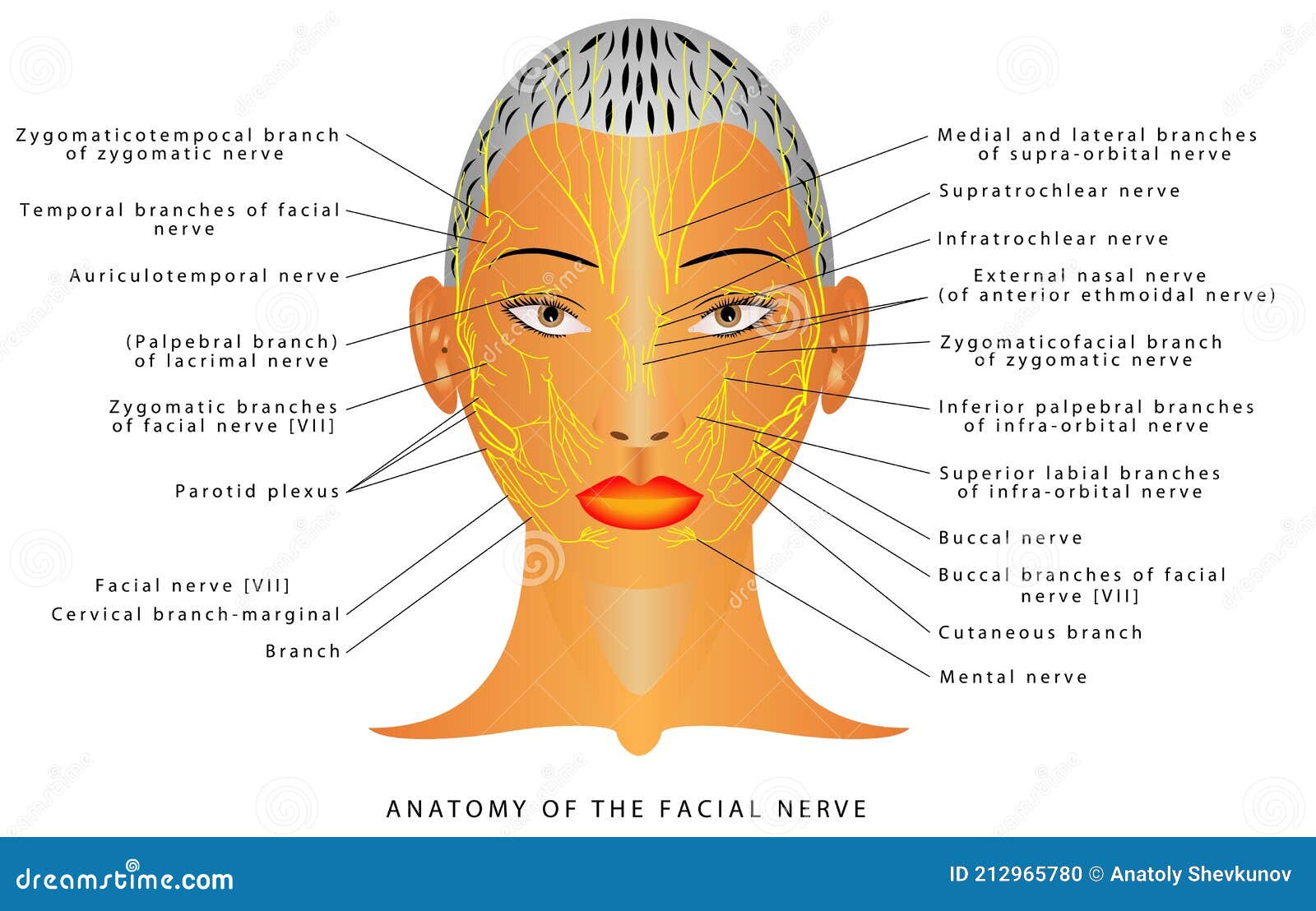
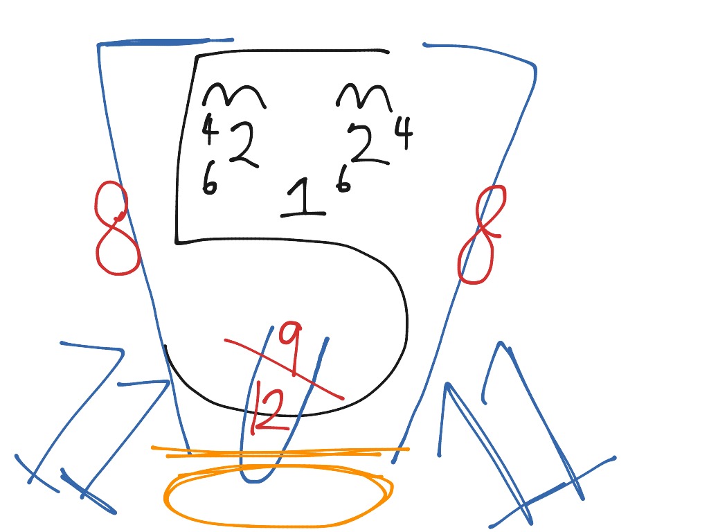
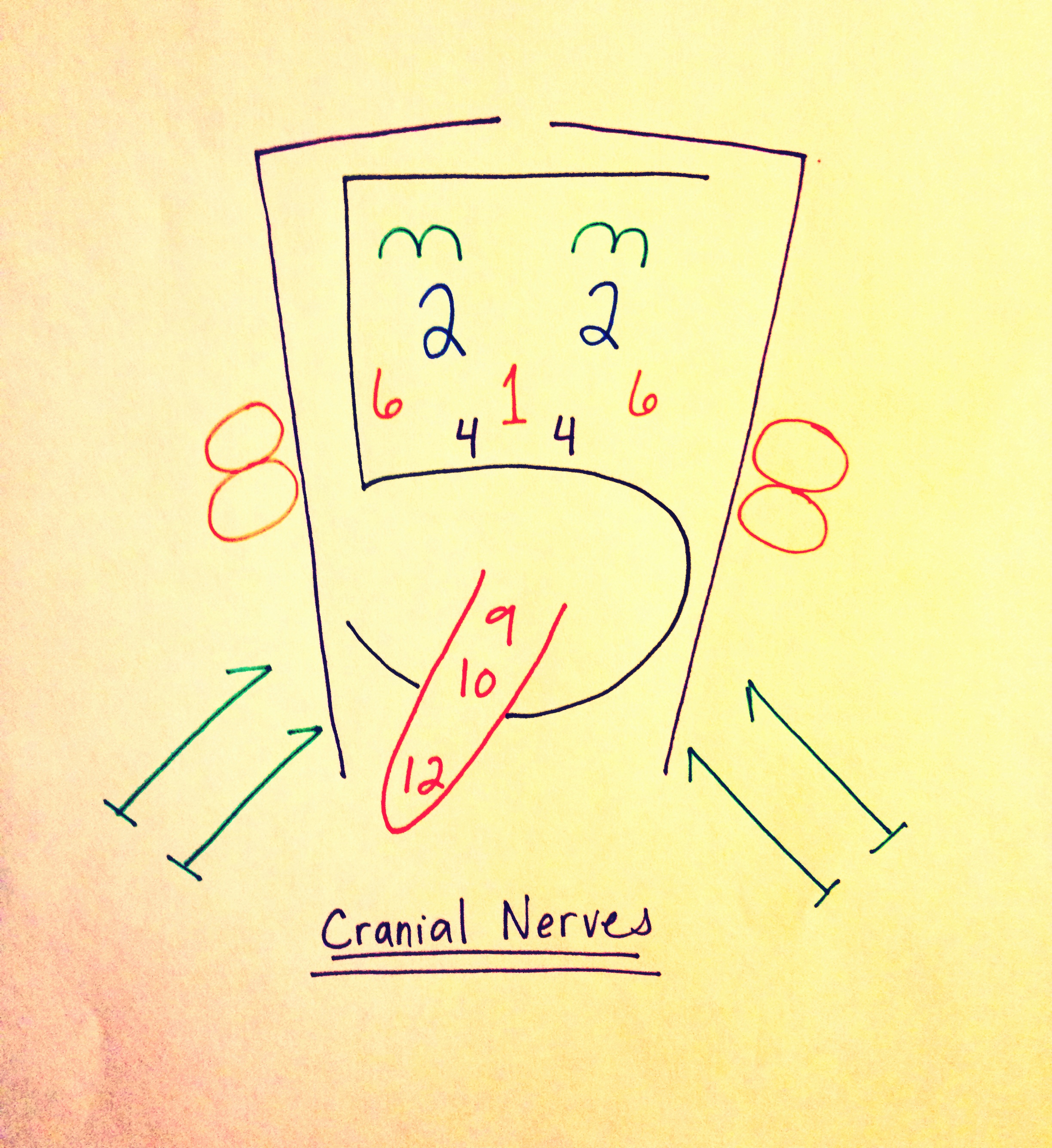
Cranial Nerves Face Drawing - This article provides a pictorial overview of the imaging of cranial nerves, with a special focus on their anatomy and pathology. Web the pattern of the facial nerve (vii) is divided into two parts, one intrapetrous part with the geniculate ganglion, the greater petrosal nerve and chorda tympani, the other part consisting of the facial area after emergence from the stylomastoid foramen. Web the facial nerve plays a key role in making facial expressions. Web from frowning to smiling, the cranial nerves help you move the muscles of your face, to even special senses such as sight and smell. Check sensation of the scalp, too. It originates in the pontomedullary region, passes through the internal auditory meatus and exits through the stylomastoid foramen. In this article, we shall look at the anatomical course of the nerve, and the motor, sensory and parasympathetic functions of its terminal branches. Web facial nerve (cn vii) cn vii is the facial nerve. Cranial nerves with the number 2 in them (e.g. Web in this video i will go over cranial nerves i through twelve by drawing a picture to help you remember. These 12 nerves control facial and eye movements and sensation. This article provides a pictorial overview of the imaging of cranial nerves, with a special focus on their anatomy and pathology. Web easy way to remember the 12 pairs of cranial nerves. Web the facial nerve, as its name suggests, controls the muscles of facial expression. The anatomy of the cranial nerves. Lower motor neurone facial nerve lesions cause upper and lower facial paralysis They control everything from your facial expression to digestion. Web the facial nerve is the seventh paired cranial nerve. Cranial nerves with the number 2 in them (e.g. Web this (much shorter) video is best watched after you've got a good grasp on the trigeminal nerve from the previous video. Each number represents one of the 12 cranial nerves, and the placement of the numbers represents the location of or an association with them. Web facial nerve (cn vii) cranial nerve 7 is a multimodal nerve, carrying both general and special fibers. Web enroll in our course: These 12 nerves control facial and eye movements and sensation. Medically reviewed by. The anatomy of the cranial nerves. This video goes through the gross a. Movement of muscles that produce facial expression Lower motor neurone facial nerve lesions cause upper and lower facial paralysis Web the facial nerve also has both motor and sensory functions. This nerve performs two major functions. It originates from the brainstem as two separate divisions; Medically reviewed by lissette pichardo, md. The facial nerve (cn vii) and the glossopharyngeal nerve (cn ix). Web facial nerve (cn vii) cranial nerve 7 is a multimodal nerve, carrying both general and special fibers. Web the facial nerve, cn vii, is the seventh paired cranial nerve. This video goes through the gross a. Lower motor neurone facial nerve lesions cause upper and lower facial paralysis Web the human body has 12 pairs of cranial nerves that control motor and sensory functions of the head and neck. It originates from the brainstem as two separate. Updated on may 31, 2023. Check sensation of the scalp, too. The facial nerve (cn vii) and the glossopharyngeal nerve (cn ix). Medically reviewed by lissette pichardo, md. Web a few years ago, a colleague taught me a much easier way to remember the cranial nerves and their locations—by drawing a face and using numbers as the facial features. It conveys some sensory information from the tongue and the interior of the. It originates from the brainstem as two separate divisions; The facial nerve is consists of four nuclei that serve different functions: Web the 12 cranial nerves are pairs of nerves that start in different parts of your brain. Your cranial nerves help you taste, smell, hear and. Web the pattern of the facial nerve (vii) is divided into two parts, one intrapetrous part with the geniculate ganglion, the greater petrosal nerve and chorda tympani, the other part consisting of the facial area after emergence from the stylomastoid foramen. Medically reviewed by lissette pichardo, md. Cranial nerves with the number 2 in them (e.g. Updated on may 31,. Web the human body has 12 pairs of cranial nerves that control motor and sensory functions of the head and neck. Updated on may 31, 2023. The facial nerve loops around the abducens nucleus. Web enroll in our course: In this article, we shall look at the anatomical course of the nerve, and the motor, sensory and parasympathetic functions of. The facial nerve loops around the abducens nucleus. Web cranial nerves send electrical signals between your brain, face, neck and torso. Web the pattern of the facial nerve (vii) is divided into two parts, one intrapetrous part with the geniculate ganglion, the greater petrosal nerve and chorda tympani, the other part consisting of the facial area after emergence from the. In this article, we shall look at the anatomical course of the nerve, and the motor, sensory and parasympathetic functions of its terminal branches. It conveys some sensory information from the tongue and the interior of the. The facial nerve is consists of four nuclei that serve different functions: Web the facial nerve, cn vii, is the seventh paired cranial. This nerve performs two major functions. Web the facial nerve plays a key role in making facial expressions. These 12 nerves control facial and eye movements and sensation. They control everything from your facial expression to digestion. It conveys some sensory information from the tongue and the interior of the. Medically reviewed by lissette pichardo, md. Updated on may 31, 2023. This article provides a pictorial overview of the imaging of cranial nerves, with a special focus on their anatomy and pathology. Web a few years ago, a colleague taught me a much easier way to remember the cranial nerves and their locations—by drawing a face and using numbers as the facial features. Web easy way to remember the 12 pairs of cranial nerves. This video goes through the gross a. Web the human body has 12 pairs of cranial nerves that control motor and sensory functions of the head and neck. Web the facial nerve, as its name suggests, controls the muscles of facial expression. Web from frowning to smiling, the cranial nerves help you move the muscles of your face, to even special senses such as sight and smell. Web the facial nerve also has both motor and sensory functions. Web the pattern of the facial nerve (vii) is divided into two parts, one intrapetrous part with the geniculate ganglion, the greater petrosal nerve and chorda tympani, the other part consisting of the facial area after emergence from the stylomastoid foramen.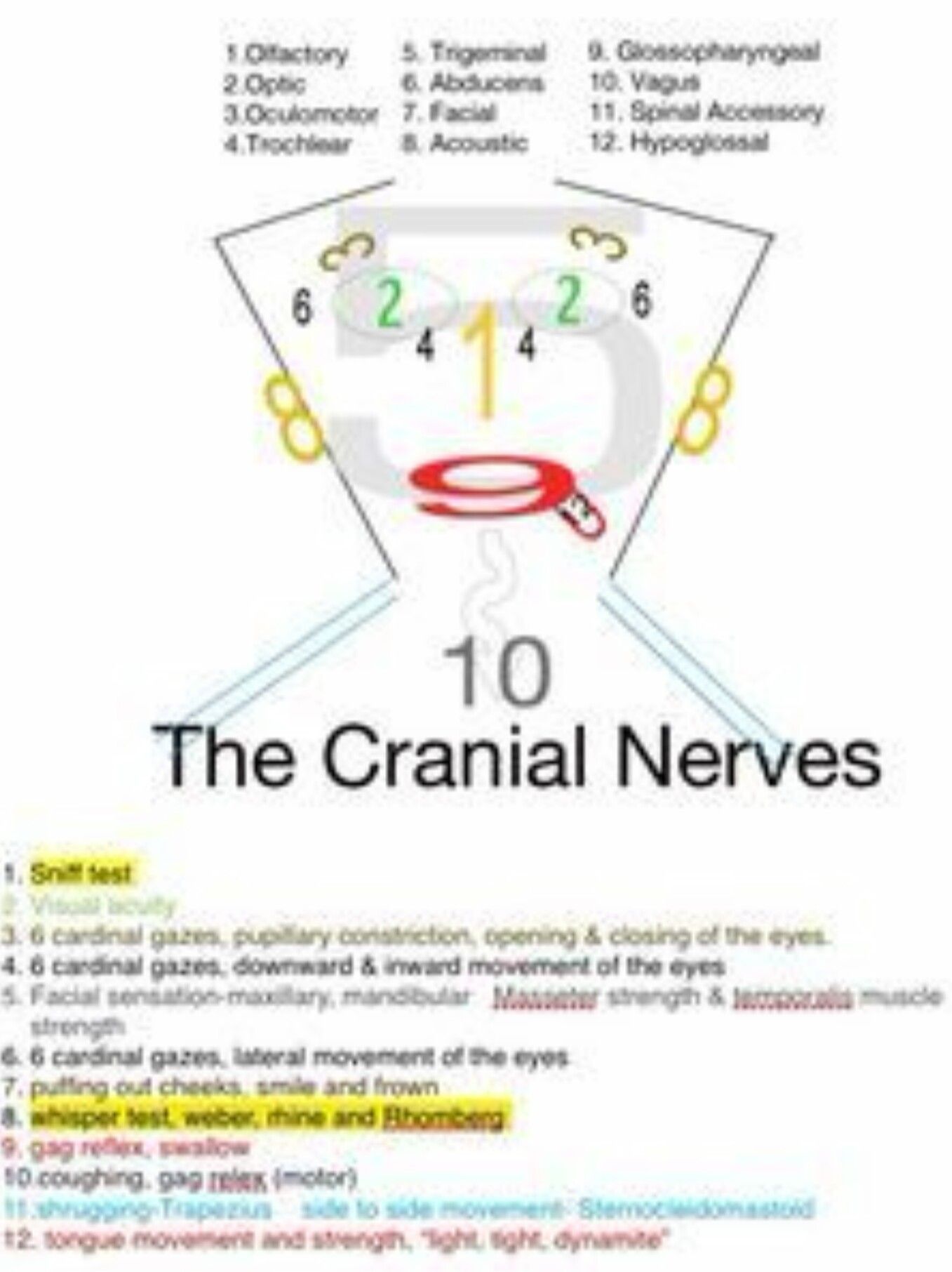
Cranial Nerve Face Drawing With Numbers at GetDrawings Free download
Cranial Nerves Face Drawing

Drawing Of The Face And Cranial Nerves 1000+ Images About Nursing
Cranial Nerve Face Drawing With Numbers at GetDrawings Free download

Cranial Nerves Drawing Face Drawing.rjuuc.edu.np

Cranial Nerve Drawing at GetDrawings Free download
Cranial Nerve Drawing at GetDrawings Free download
Cranial Nerves Face

Cranial Nerve Face Drawing With Numbers at GetDrawings Free download
Cranial Nerve Face Drawing With Numbers at GetDrawings Free download
Which Cranial Nerves Are Involved In Taste?
This Video Is An Overview Of The Function Of The Following Cranial Nerves:1.
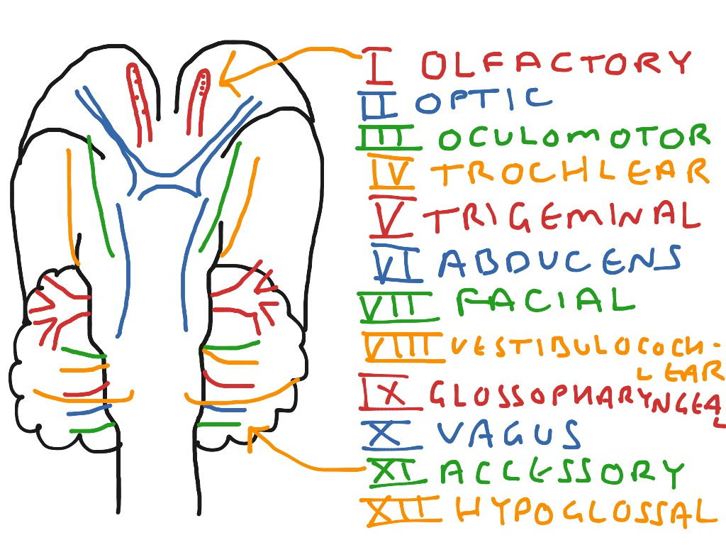
The Location Of The Cranial Nerves On The Cerebrum And Brainstem.
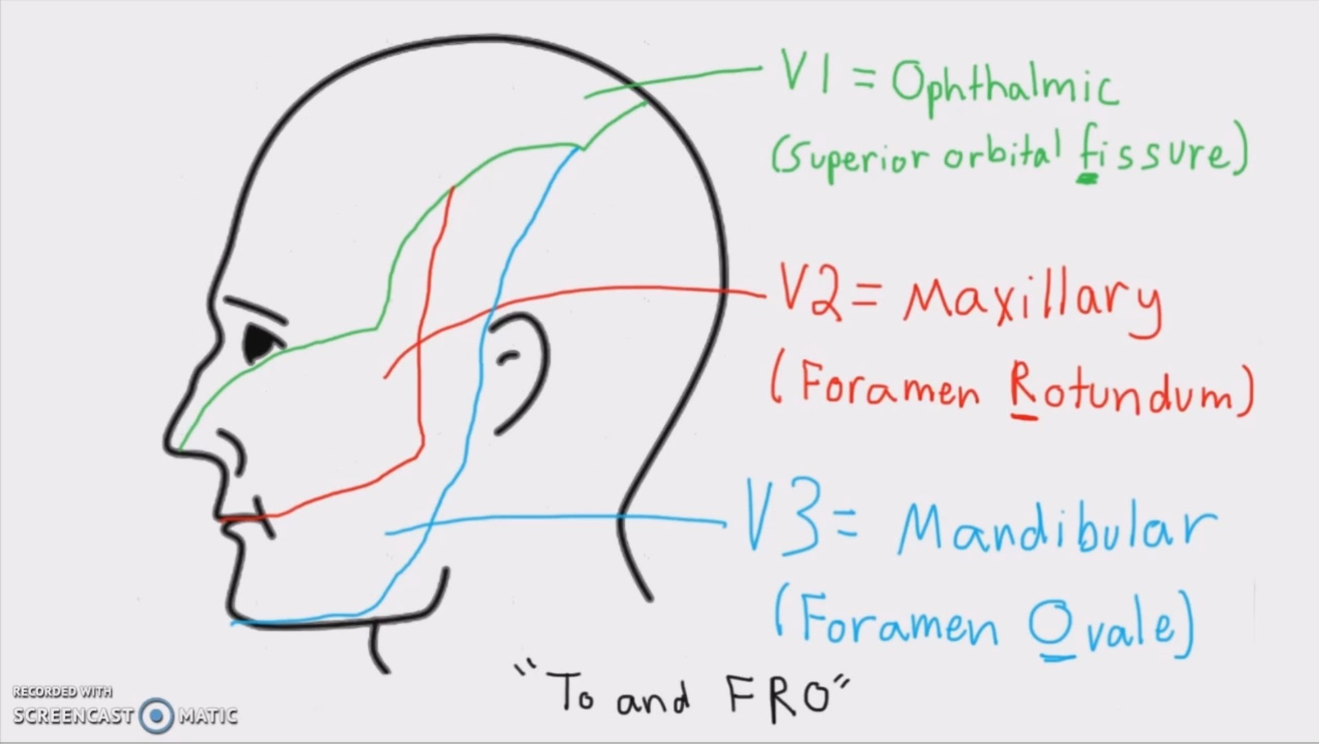
Web This (Much Shorter) Video Is Best Watched After You've Got A Good Grasp On The Trigeminal Nerve From The Previous Video.
Related Post: