Drawing Of The Brain With Labels
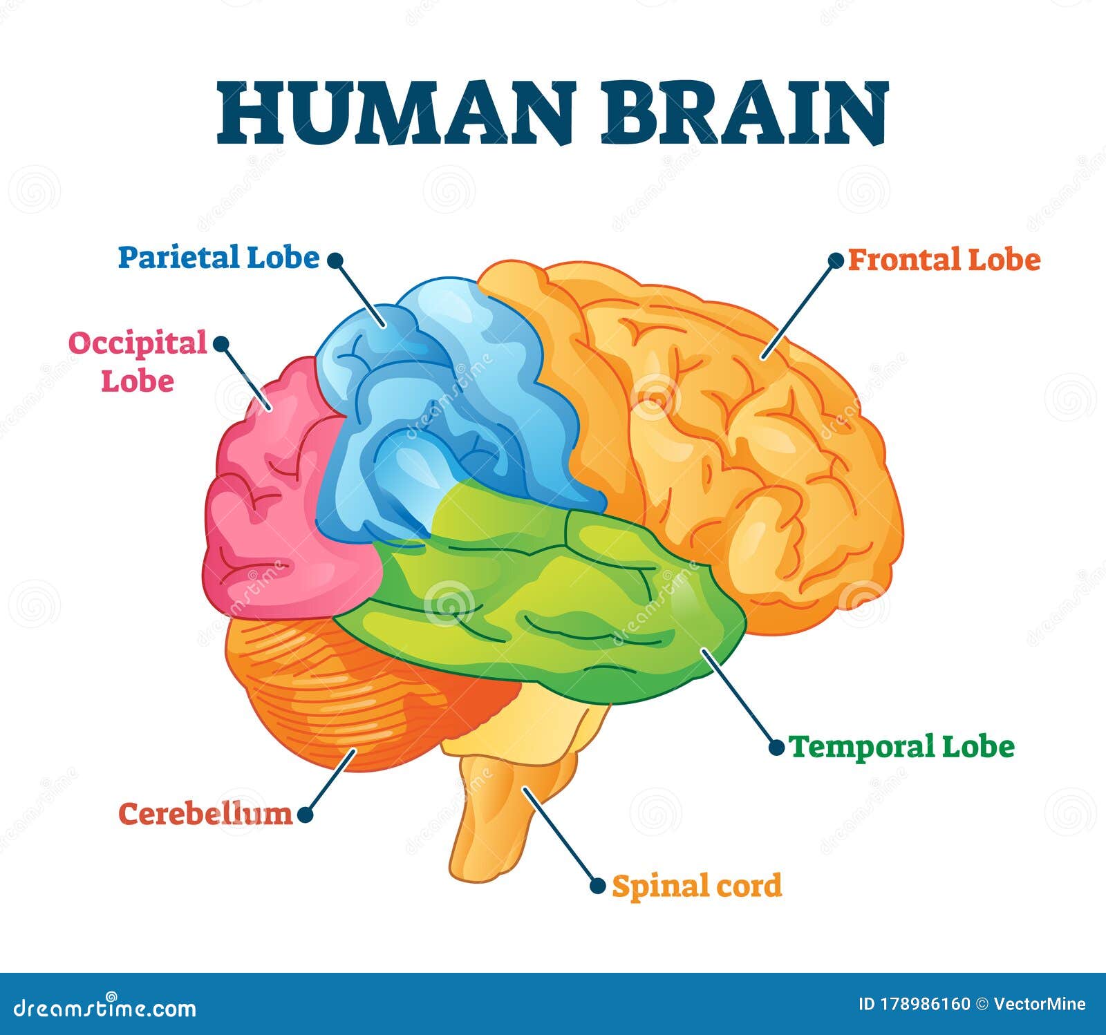
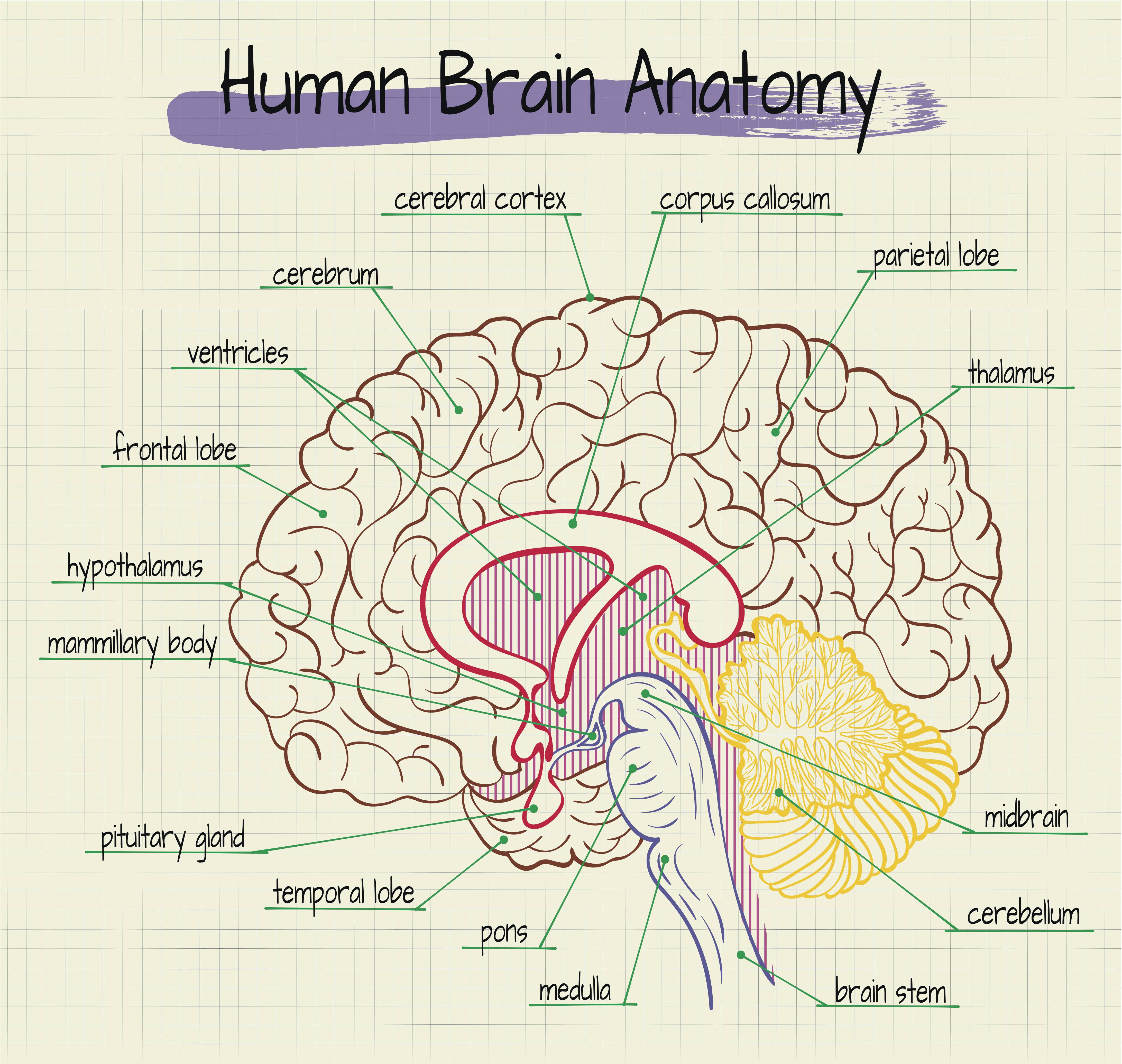
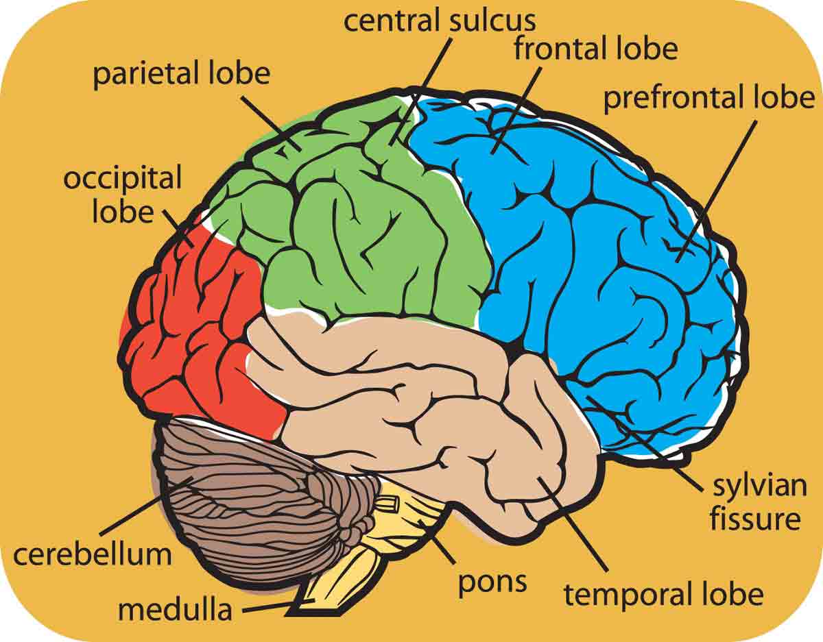
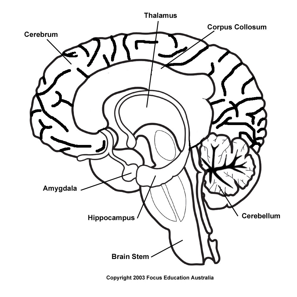
Drawing Of The Brain With Labels - Try to memorize the name and location of each structure, then proceed to test yourself with the blank brain diagram provided below. Each hemisphere is conventionally divided into six lobes, but only four of them are visible from this lateral perspective.the lobes are. A lateral view of the cerebrum is the best perspective to appreciate the lobes of the hemispheres. Web labeled brain diagram. Web the cerebellum adjusts body movements, speech coordination, and balance, while the brain stem relays signals from the spinal cord and directs basic internal functions and reflexes. The first version is color coded by section. The second version is the natural color of the human brain, and the third. Web brain cells can be broken into two groups: Use of interactive anatomical labels. The occipital lobe is the back part of the brain that is involved with vision. You’ll be on your way to mastering how to draw a brain. The frontal lobe, parietal lobe, temporal lobe, occipital lobe, cerebellum, and brainstem. Web the human brain consists of several parts which are clearly labeled in the video. Most of the neurons in the. The diagram is available in 3 versions. Web the parietal lobe houses wernicke’s area, which helps the brain understand spoken language. The second version is the natural color of the human brain, and the third. The cerebrum, the largest part, is responsible for sensory interpretation, thought processing, and voluntary muscle activity. High intellectual functions occur in the cerebrum. The brainstem connects the brain. The labeled human brain diagram contains labels for: How to view anatomical labels. It provides access to an atlas and to images in axial planes, allowing the user to learn and review neuroanatomy. Web free download 45 best quality drawing of the brain with labels at getdrawings. It also functions as the coordinating centre of intellectual, sensation and the nervous. 85% of the brain is cerebral cortex, divided as, 41% frontal lobe, 22% temporal lobe, 19% parietal lobe and 18% occipital lobe. Web the human brain consists of several parts which are clearly labeled in the video. High intellectual functions occur in the cerebrum. The brain is an organ made up of neural tissue. Instead of coloring and labeling on. Web there are only a few creatures without having a brain. Use of interactive anatomical labels. If you’re freaking out thinking drawing a brain is like climbing mount everest, chill. The brain is an organ made up of neural tissue. The frontal lobe, parietal lobe, temporal lobe, occipital lobe, cerebellum, and brainstem. Web labeled brain diagram. Use of interactive anatomical labels. Web the human brain is a complex organ, made up of several distinct parts, each responsible for different functions. First up, have a look at the labeled brain structures on the image below. Each hemisphere is conventionally divided into six lobes, but only four of them are visible from this lateral. Forming the brain with a pencil sketch; Web brain cells can be broken into two groups: Web labeled human brain diagram. How to view anatomical labels. Web free download 45 best quality drawing of the brain with labels at getdrawings. Web atlas of the human brain based on colored anatomical drawings and diagrams. To draw an anatomically accurate brain, draw a curve in the shape of the lengthwise half of a large egg, making the right side more curved. Neurons, or nerve cells, are the cells that perform all of the communication and processing within the brain. Web labeled brain. Outlining intricate parts of the brain; Web free download 45 best quality drawing of the brain with labels at getdrawings. High intellectual functions occur in the cerebrum. The occipital lobe is the back part of the brain that is involved with vision. How to view anatomical labels. The occipital lobe is the back part of the brain that is involved with vision. The brainstem connects the brain. Web anatomy of the brain: If you’re freaking out thinking drawing a brain is like climbing mount everest, chill. Web this brain labeling activity was created for remote learners as an alternative to the labeling and coloring worksheet we would. Web discover the intricacies of the human brain with our labeled drawing video. The user can select to display multiple categories of labels on the illustrations: Web the human brain is a complex organ, made up of several distinct parts, each responsible for different functions. Web hi everyone, in this video i show you how to draw the human brain. In this informative tutorial, each part of the brain is clearly labeled, making it a valuable resource for studying and acing your exams. Forming the brain with a pencil sketch; Web more than half of the neurons in the brain are found in the cerebellum and only 10% neurons make up the brain. The frontal lobe, parietal lobe, temporal lobe,. The cerebrum, the largest part, is responsible for sensory interpretation, thought processing, and voluntary muscle activity. Labeled diagram showing the main parts of the brain This module is a comprehensive and affordable learning tool for medical students and residents and especially for neuroradiologists and radiation oncologists. Web hi everyone, in this video i show you how to draw the human brain step by step 🧠. The brain is an organ made up of neural tissue. Use of interactive anatomical labels. It provides access to an atlas and to images in axial planes, allowing the user to learn and review neuroanatomy. The diagram of the brain is useful for both class 10 and 12. 85% of the brain is cerebral cortex, divided as, 41% frontal lobe, 22% temporal lobe, 19% parietal lobe and 18% occipital lobe. Enhance your understanding of the brain's structure and improve your chances of scoring higher grades. Try to memorize the name and location of each structure, then proceed to test yourself with the blank brain diagram provided below. The occipital lobe is the back part of the brain that is involved with vision. It’s like a map for your brain drawing journey. This will help with your exams and can score higher marks. Neurons, or nerve cells, are the cells that perform all of the communication and processing within the brain. First up, have a look at the labeled brain structures on the image below.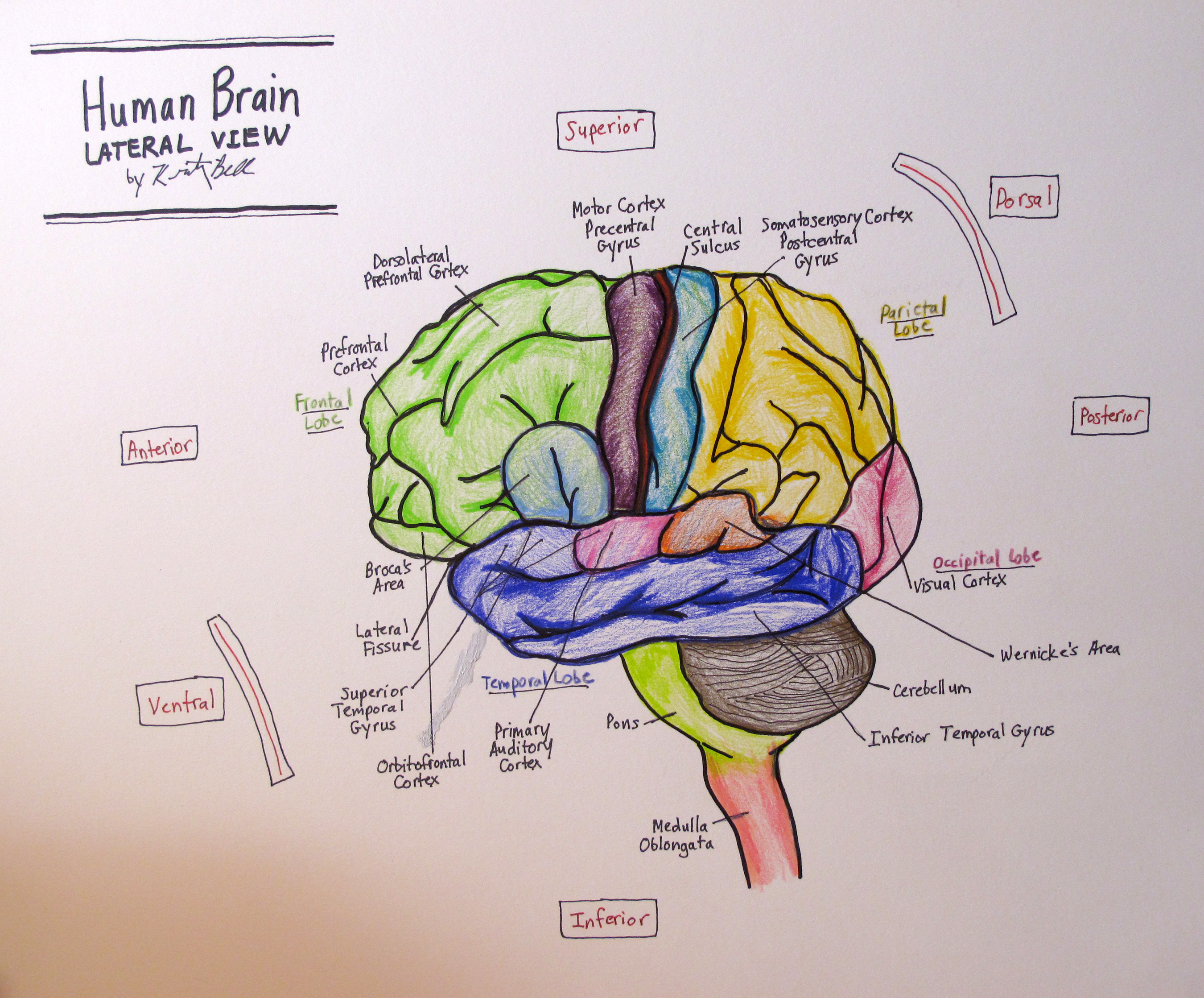
Brain Drawing With Labels at GetDrawings Free download

Brain Images Labeled

Brain Diagram Labeled Tim's Printables
Brain Parts Stock Illustrations 7,257 Brain Parts Stock Illustrations
Labeled Diagram Of A Brain

How to Draw a Brain 14 Steps wikiHow
Brain Drawing With Labels at GetDrawings Free download
Drawing Of The Brain With Labels at Explore

Pin on Education

Brain Map 2
Web Explore The Structure And Function Of The Human Brain In 3D, With Interactive Models, Videos, And Articles On Brainfacts.org.
Web Table Of Contents.
The First Version Is Color Coded By Section.
The Frontal Lobe, Parietal Lobe, Temporal Lobe, Occipital Lobe, Cerebellum, And Brainstem.
Related Post: